Home
Uncategories
Chest Muscles Diagram : How Many Muscles Are In The Human Body Plus A Diagram : The pectoralis major muscles (also known as the pecs) are located on the front of the rib cage.
Chest Muscles Diagram : How Many Muscles Are In The Human Body Plus A Diagram : The pectoralis major muscles (also known as the pecs) are located on the front of the rib cage.
Chest Muscles Diagram : How Many Muscles Are In The Human Body Plus A Diagram : The pectoralis major muscles (also known as the pecs) are located on the front of the rib cage.. The chest is the area of origin for many of the body's systems as it houses organs such as the heart, esophagus, trachea, lungs, and thoracic diaphragm. If you are pulling your muscles more than enough and doing burdening lots of effort, then the pain that is going to felt by you is no the symptom of the muscular chest pain. These important muscles control many motions that involve moving the arms and head — such as throwing a ball, looking up at the sky, and raising your hand. The dominant muscle in the upper chest is the pectoralis major. Muscles allow a person to move, speak, and chew.
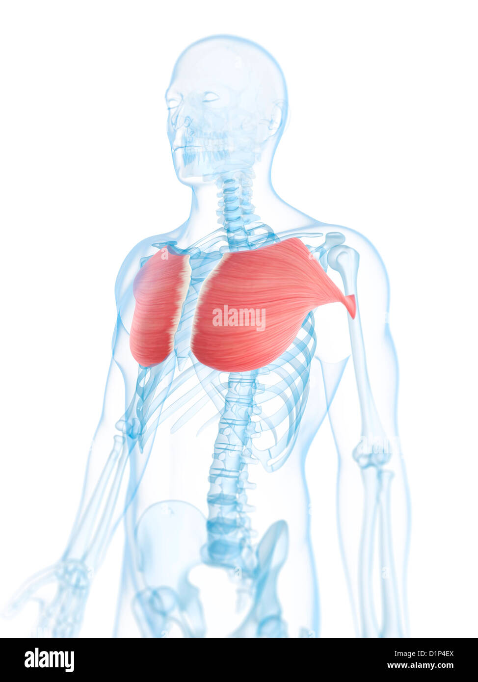
The pecs attach to the humerus near the shoulder joint and originate on the breastbone in the center of the chest. Pectoral muscles (colloquially referred to as pecs) are the muscles that connect the front of the human chest with the bones of the upper arm and shoulder. The pectoralis major, pectoralis minor, serratus anterior and subclavius. The usual cause of overstretched chest muscles can be the over exercising. Muscles in chest area human chest muscles pectoral.
Chest Muscles Artwork Stock Photo Alamy from c8.alamy.com The pectoralis major muscles (also known as the pecs) are located on the front of the rib cage. These important muscles control many motions that involve moving the arms and head — such as throwing a ball, looking up at the sky, and raising your hand. Doctors diagnose chest wall pain in at least 25% of patients who come to the emergency room for chest pain. This page provides an overview of the chest muscle group. The suboccipital muscles act to rotate the head and extend the neck. Related posts of chest muscles diagram. The chest is part of a larger group of pushing muscles found in the upper body. Muscles allow a person to move, speak, and chew.
Chest muscle anatomy diagram / located immediately below the skin) muscles of the body.
Related posts of chest muscles diagram anatomy muscle arm. Human chest muscles diagram : The pectoralis major muscles (also known as the pecs) are located on the front of the rib cage. During bench press, large forces the muscle is required to generate to lift and lower the bar, combined with an overstretching of the muscle, can place too much stress on the. The dominant muscle in the upper chest is the pectoralis major. Anatomy of the chest muscles. Muscles of the hip, thigh, and leg. The levator scapulae muscle is located near the neck, around the neck's base and side. Several muscles that move the arms, head, and neck have their origins on the sternum. 1.upper chest 2.middle chest 3.lower chest 4.interior chest 5.outer chest. Chest muscles are responsible for adduction, internal rotation, and forwards flexion of the humerus. The pectoral region is located on the anterior chest wall. Almost every muscle constitutes one part of a pair of identical.
This page provides an overview of the chest muscle group. The chest muscles are made up of the pectoralis major and, underneath that, the pectoralis minor. The pectoral region is located on the anterior chest wall. The pectoralis major muscles (also known as the pecs) are located on the front of the rib cage. If you are pulling your muscles more than enough and doing burdening lots of effort, then the pain that is going to felt by you is no the symptom of the muscular chest pain.
Chest Anatomy What Are The Muscles And What Do They Do Openfit from cdn.prod.openfit.com Chest muscle anatomy diagram / located immediately below the skin) muscles of the body. Neck and chest muscles diagram neck and chest muscles diagram : Chest muscles anatomy for bodybuilders. To fully develop your chest, you need to hit it with heavy weight using a couple smartly chosen exercises. I often get asked, how can i build thick powerful pecs? Your pectoralis major and pectoralis minor muscles make up most of the muscle mass in your chest. Almost every muscle constitutes one part of a pair of identical. Anatomy of the chest and the lungs:
Human chest muscles graph diagram from graphdiagram.com small muscles running between the ribs, known as the external intercostal muscles, lift the ribs during deep breathing to further expand the chest and lungs and provide even more air to the body.
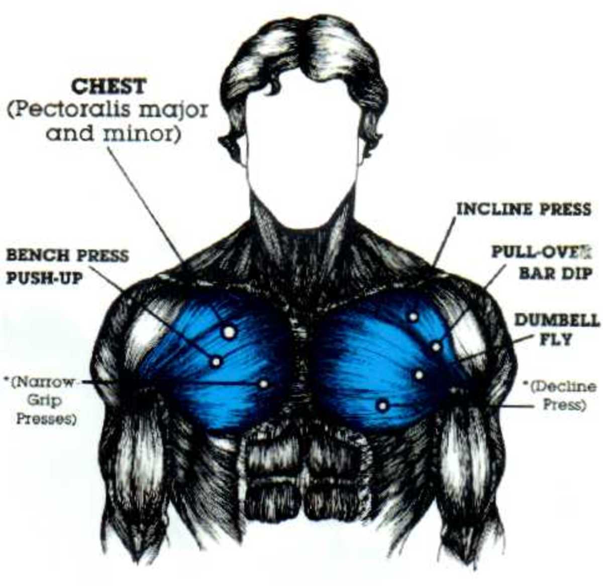
The dominant muscle in the upper chest is the pectoralis major. The muscles of the chest and upper back occupy the thoracic region of the body inferior to the neck and superior to the abdominal region and include the muscles of the shoulders. Related posts of chest muscles diagram. Your pectoralis major and pectoralis minor muscles make up most of the muscle mass in your chest. The chest muscles of our body can get pulled and strained. The primary muscle in the chest region is called the pectoralis major. Abdominal muscles diagram, back muscles diagram, chest muscle diagram exercise, chest workout diagram, female pectoral muscles, female pelvic muscles diagram, shoulder muscles diagram, womens chest muscles, human muscles, abdominal muscles diagram, back muscles diagram, chest muscle diagram. The pecs attach to the humerus near the shoulder joint and originate on the breastbone in the center of the chest. These important muscles control many motions that involve moving the arms and head — such as throwing a ball, looking up at the sky, and raising your hand. Anatomy muscle arm 12 photos of the anatomy muscle arm anatomy muscle arm quiz, anatomy muscles of the. It contains four muscles that exert a force on the upper limb: Chest muscles anatomy for bodybuilders. To fully develop your chest, you need to hit it with heavy weight using a couple smartly chosen exercises.
The pectoralis major, pectoralis minor, serratus anterior and subclavius. Elbow muscle anatomy mri 12 photos of the elbow muscle anatomy mri elbow muscle. The chest exercises and workouts you need to build bigger pecs. The chest is part of a larger group of pushing muscles found in the upper body. Anatomy of the chest muscles.
Developing Those Chest Muscles Caloriebee from images.saymedia-content.com The fibers of the pectoralis muscles run like a fan across the chest. Learn vocabulary, terms and more with flashcards, games and other study tools. The pecs attach to the humerus near the shoulder joint and originate on the breastbone in the center of the chest. The chest is part of a larger group of pushing muscles found in the upper body. The levator scapulae muscle is located near the neck, around the neck's base and side. Unfortunately, in many cases, that's as far as the doctor takes the diagnosis. The dominant muscle in the upper chest is the pectoralis major. It contains four muscles that exert a force on the upper limb:
These important muscles control many motions that involve moving the arms and head — such as throwing a ball, looking up at the sky, and raising your hand.
These important muscles control many motions that involve moving the arms and head — such as throwing a ball, looking up at the sky, and raising your hand. In this article, we shall learn about the anatomy of the muscles of the anterior chest. Chest muscle anatomy the pectoralis major muscles also known as the pecs are located on the front of the rib cage and form the major muscles of the chest. 1.upper chest 2.middle chest 3.lower chest 4.interior chest 5.outer chest. Anatomy of the chest and the lungs: The shoulder muscles bridge the transitions from the torso into the head/neck area and into the uppe. The pectoralis major, pectoralis minor, serratus anterior and subclavius. The pectoral region is located on the anterior chest wall. Classic symptoms of strain in the chest muscle include: Chest muscles, chest muscle diagram. Primarily, there are three chest muscles involved in movement: These do not include the hip, neck and forearm muscles, which are rarely trained in isolation. Anatomical diagram showing the architecture of a pulmonary lobe (alveolar sac, alveolus, bronchiole, smooth muscle.) almost every muscle constitutes one part of a pair of identical bilateral muscles, found on both sides, resulting in approximately 320 pairs of muscles.

0 Comments:
Posting Komentar